Rib Cage Anatomy - Human Ribs Diagramnumbered - The scalene muscles help with neck flexion and side bending.
byAdmin•
0
Rib Cage Anatomy - Human Ribs Diagramnumbered - The scalene muscles help with neck flexion and side bending.. It has a roughened area on its upper surface, from which the serratus anterior muscle originates. Bifid ribs occur in up to 8.4% of samoans. It is supported by the vertical sternum or. Nov 18, 2020 · exhale and allow your rib cage and upper back come back to their natural position. In the mammary line (an imaginary vertical line from the right breast nipple), the liver extends from the 5th rib to the bottom of the rib cage 9.
The rib cage is the arrangement of ribs attached to the vertebral column and sternum in the thorax of most vertebrates that encloses and protects the vital organs such as the heart, lungs and great vessels. Nov 18, 2020 · exhale and allow your rib cage and upper back come back to their natural position. In the mammary line (an imaginary vertical line from the right breast nipple), the liver extends from the 5th rib to the bottom of the rib cage 9. The anterior and middle scalenes attach to the first rib, while the posterior scalene attaches to the second rib. The remaining ribs are called floating ribs and only attach to the spine.
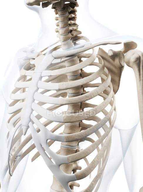
Human rib cage anatomy — human anatomy, 3d - Stock Photo ... from st.focusedcollection.com The human rib cage is a component of the human respiratory system. The scalene muscles are 3 pairs of lateral neck muscles that connect the mid and lower cervical spine with the top of the rib cage. Hepar (in ancient greek) = liver In the mammary line (an imaginary vertical line from the right breast nipple), the liver extends from the 5th rib to the bottom of the rib cage 9. Sep 06, 2019 · there are 11 pairs of external intercostal muscles. Learn anatomy with free interactive flashcards. The sternal end of the rib is cleaved into two. There are 12 pairs of ribs, and the first 10 articulate with both the thoracic spine and the costal cartilage on the front of the rib cage.
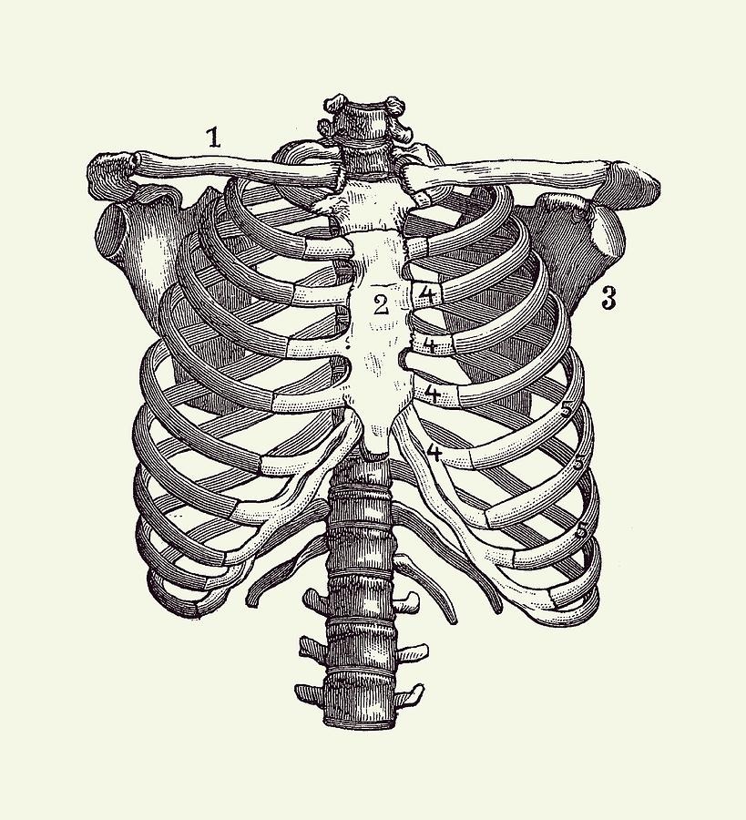
It is formed by the 12 thoracic vertebrae, 12 pairs of ribs and associated costal cartilages and the sternum.
Learn anatomy with free interactive flashcards. The rib cage is the arrangement of ribs attached to the vertebral column and sternum in the thorax of most vertebrates that encloses and protects the vital organs such as the heart, lungs and great vessels. The scalene muscles are 3 pairs of lateral neck muscles that connect the mid and lower cervical spine with the top of the rib cage. The scalene muscles help with neck flexion and side bending. The human rib cage is a component of the human respiratory system. In the mammary line (an imaginary vertical line from the right breast nipple), the liver extends from the 5th rib to the bottom of the rib cage 9. A bifid rib is a congenital abnormality of the rib cage and associated muscles and nerves which occurs in about 1.2% of humans. They run inferoanteriorly from the rib above to the rib below, and are continuous with the external oblique of the abdomen. The remaining ribs are called floating ribs and only attach to the spine. Jul 27, 2021 · the thoracic cage (rib cage) is the skeleton of the thoracic wall. Since the ribs and rib cage are attached to the spine, dysfunction in the spine can cause symptoms and problems in the ribs. The sternal end of the rib is cleaved into two. The thoracic cage takes the form of a domed bird cage with the horizontal bars formed by ribs and costal cartilages.
Learn anatomy with free interactive flashcards. The scalene muscles are 3 pairs of lateral neck muscles that connect the mid and lower cervical spine with the top of the rib cage. Jul 27, 2021 · the thoracic cage (rib cage) is the skeleton of the thoracic wall. The remaining ribs are called floating ribs and only attach to the spine. Nov 18, 2020 · exhale and allow your rib cage and upper back come back to their natural position.
Human Heart Inside Rib Cage Dictionary Art Print No. 9 ... from i.pinimg.com Originate at the lower border of the rib, inserting into the superior border of the rib below. Bifid ribs occur in up to 8.4% of samoans. It encloses the thoracic cavity, which contains the lungs. You may find that with practice, this natural, familiar, habitual position changes, and you acquire more distance between your ribs and pelvis. The remaining ribs are called floating ribs and only attach to the spine. Elevates the ribs, increasing the thoracic volume. Rib 2 is thinner and longer than rib 1, and has two articular facets on the head as normal. Jul 27, 2021 · the thoracic cage (rib cage) is the skeleton of the thoracic wall.
The scalene muscles are 3 pairs of lateral neck muscles that connect the mid and lower cervical spine with the top of the rib cage.
You may find that with practice, this natural, familiar, habitual position changes, and you acquire more distance between your ribs and pelvis. There are 12 pairs of ribs, and the first 10 articulate with both the thoracic spine and the costal cartilage on the front of the rib cage. A bifid rib is a congenital abnormality of the rib cage and associated muscles and nerves which occurs in about 1.2% of humans. Elevates the ribs, increasing the thoracic volume. The human rib cage is a component of the human respiratory system. The remaining ribs are called floating ribs and only attach to the spine. Choose from 500 different sets of anatomy flashcards on quizlet. An inhalation is accomplished when the muscular diaphragm, at the floor of the thoracic cavity, contracts and flattens, while the contraction of intercostal muscles lift the rib cage up and out. They run inferoanteriorly from the rib above to the rib below, and are continuous with the external oblique of the abdomen. The thoracic cage takes the form of a domed bird cage with the horizontal bars formed by ribs and costal cartilages. Originate at the lower border of the rib, inserting into the superior border of the rib below. The anterior and middle scalenes attach to the first rib, while the posterior scalene attaches to the second rib. It has a roughened area on its upper surface, from which the serratus anterior muscle originates.
Jul 27, 2021 · the thoracic cage (rib cage) is the skeleton of the thoracic wall. Bifid ribs occur in up to 8.4% of samoans. They run inferoanteriorly from the rib above to the rib below, and are continuous with the external oblique of the abdomen. You may find that with practice, this natural, familiar, habitual position changes, and you acquire more distance between your ribs and pelvis. Choose from 500 different sets of anatomy flashcards on quizlet.
Shoulder and Rib Cage Diagram - Vintage Anatomy Poster 2 ... from images.fineartamerica.com It encloses the thoracic cavity, which contains the lungs. It is supported by the vertical sternum or. The scalene muscles help with neck flexion and side bending. Rib 2 is thinner and longer than rib 1, and has two articular facets on the head as normal. Since the ribs and rib cage are attached to the spine, dysfunction in the spine can cause symptoms and problems in the ribs. Jul 27, 2021 · the thoracic cage (rib cage) is the skeleton of the thoracic wall. Choose from 500 different sets of anatomy flashcards on quizlet. An inhalation is accomplished when the muscular diaphragm, at the floor of the thoracic cavity, contracts and flattens, while the contraction of intercostal muscles lift the rib cage up and out.
It encloses the thoracic cavity, which contains the lungs.
Since the ribs and rib cage are attached to the spine, dysfunction in the spine can cause symptoms and problems in the ribs. The scalene muscles are 3 pairs of lateral neck muscles that connect the mid and lower cervical spine with the top of the rib cage. Elevates the ribs, increasing the thoracic volume. Jul 27, 2021 · the thoracic cage (rib cage) is the skeleton of the thoracic wall. Hepar (in ancient greek) = liver You may find that with practice, this natural, familiar, habitual position changes, and you acquire more distance between your ribs and pelvis. An inhalation is accomplished when the muscular diaphragm, at the floor of the thoracic cavity, contracts and flattens, while the contraction of intercostal muscles lift the rib cage up and out. The human rib cage is a component of the human respiratory system. Choose from 500 different sets of anatomy flashcards on quizlet. In the mammary line (an imaginary vertical line from the right breast nipple), the liver extends from the 5th rib to the bottom of the rib cage 9. It encloses the thoracic cavity, which contains the lungs. There are 12 pairs of ribs, and the first 10 articulate with both the thoracic spine and the costal cartilage on the front of the rib cage. The scalene muscles help with neck flexion and side bending.